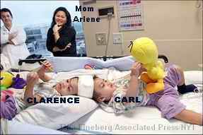
The Aguirre Twins --- A Neurosurgical Adventure!
The separation of conjoined craniopagus Aguirre twins is the first of its kind in New York. Craniopagus twins, as mentioned in my previous posts, is one of the rarest conditions a doctor might encounter in his medical practice, occurring about 1 in 10,000,000 births. Statistics-wise, craniopagus separation is a complex and dangerous neurosurgical procedure that has successfully been done fewer than 10 times, and never in New York Dr. James T. Goodrich, director of pediatric neurosurgery at The Children's Hospital at Montefiore (CHAM), and Dr. David Staffenberg, the chief of plastic surgery at Montefiore, leaders of the operating team, used a different surgical approach in the separation. They proceeded in surgical steps spread over several months, rather than one long marathon session. This was done to minimize blood loss and prevent brain swelling. Other operations to separate craniophagus twins were usually accomplished in one long, marathon sesssion like that of Egyptian twins Ahmed and Mohamed Ibrahim, which took 34 straight hours of surgery at Children's Medical Center in Dallas, last October 12, 2003. Today, both boys, are making progress in their neurologic recovery and show no signs of infection or other problems. They are an inspiration to the medical team involved with the Aguirre twins.
The CHAM Medical Team tasked to separate the twins is made up of 16 doctors, including Dr. Willy Lopez of the UP-PGH, the neurosurgeon who looked after the twins here in Manila. The rest of the team are nurses, child care specialists, and social workers.
The ultimate objective of the Aguirre craniopagus case is to produce two separate, fully functional boy siblings who will grow up independently. According to Montefiore hospital staff, if that goal comes into reality, it will be the first time conjoined twins have survived without at least one suffering some brain damage.
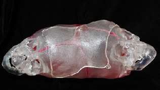
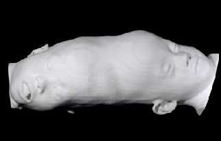
The neurosurgeons planned their approach using anatomical models made by Medical Modelling, a Colorado company that also provided similar models to the doctors who separated the Egyptian twin boys in Dallas. The models are so exquisitely done they provide 3D-images and models of the boys' brains, and are very useful in surgical planning. The company used an advanced computer software to view and extract information from CT and MRI images of Clarence and Carl's skull structures, so the models produced are precise and almost like the actual skulls. Most of the images I used here to illustrate the twins' condition during the surgical procedures came from Medical Modelling.
Here are the series of neurosurgeries that led to the separation of twins Clarence and Carl:
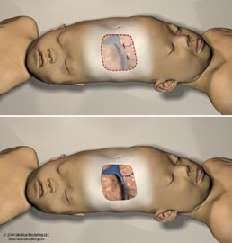
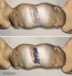
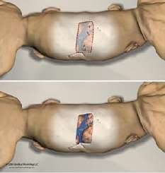
- October 20,2003 - a 5-hour operation that was like a probe mission to explore the twins' brains and see if there were any overlooked structure not represented by the anatomic models mentioned. The doctors performed a craniotomy, in which they removed a small piece of skull on the twins to provide a window in which to work. Dr. David Staffenberg then implanted deflated balloons under the twins' scalp, which are actually tissue expanders. Tissue expanders were filled with sterile saline through a port to stretch the overlying skin. Over time, the doctors will need the "stretched skin" provided by the tissue expanders to cover the exposed brain tissue of the twins once they are separated.
- November 24, 2003 - 5½-hour operation that divided the blood vessels shared by the twins. A new set of tissue expanders were inserted under the skin, again inflated with saline to stretch the skin and create more of it. The craniotomy done this time was more toward the back of their heads than the first one. Four blood vessels were separated, including part of the sagittal sinus, which is a major vein of the brain. It was during this operation when the doctors found a surprise: Clarence, who was the smaller twin, had a more complete system of veins. Initially, doctors had the impression that it was Carl who had that distinction. Dr. Lopez and Dr. Goodrich also separated a section of the twins' brains by pushing them apart. Dr. Goodrich said that the twins' brains abutted each other but were not intertwined.
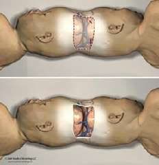
- February 20, 2004 - another 5-hour operation which was described as "more treacherous than expected." Part of the challenge was the difficulty encountered in removing a skull bone because it was not only stuck to the membrane surrounding the brain, but had veins running through it. Dr. Goodrich said it was difficult because dealing with the said veins was like working on something that had the "consistency of wet toilet paper." And it was during this stage of the operation "when things go badly, they go very, very badly and when they go well, they go extremely well." It was good that the medical team encountered more of the latter than the former. Doctors continued gently pushing apart the two brains of the twins and a thin plastic sheet was inserted to keep them apart. In this operation they gave Clarence both a part of the sagittal sinus and the pool of blood vessels that had formed between the two brains. The doctors had decided that Clarence naturally owned the major part of the veins, and they wanted Carl to continue developing a secondary vein system, something he started to do after the November surgery.
- August 4, 2004 - a 17-hour operation that finally separated the twins. Ten hours into the surgery, the doctors discovered another surprise: the twins' brains were actually fused! In the initial operations, they thought that the brains "abutted each other but were not intertwined." Now they discovered they were wrong as they found that the twins' brains were fused at a 2-inch-square section close to the top of their heads. After about 90 minutes of soul-searching, the medical team decided to proceed and separate the brains. After the gruelling operation, Clarence and Carl lay side by side, each in his own bed, for the first time in their lives.
[References: Melissa Klein and Laura Babcock of TheJournalNews.com, New York Times]
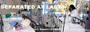
Today the two Filipino twins are sharing a room in Montefiore's pediatric intensive care unit, sedated, on ventilators, and recovering in side-by-side beds. They are sedated mostly for pain management and to allow recovery by preventing both from making unnecessary movements after the surgery. They are recovering well according to Dr. Robert W. Marion, the twins' pediatrician, and have stable vital signs except for a slightly high blood pressure for Clarence. Yesterday, a CT scan was done on Carl Aguirre. Doctors speculate that Carl might need a shunt later on to help drain fluids from around his brain, since Clarence retained most of the veins that would do that. Dr. Staffenberg also changed the twins' bandages yesterday. So far, there were no signs of infection and brain swelling noted.
The fourth operation is not the last. The twins are still going to undergo several more operations in the future, most of them reconstructive. At this point, the twins need our prayers. Let us also pray for the doctors who will be doing more surgical operations on the two in the months to come.
It should also be noted that the doctors still do not know the effects of separating the fused brain sections of Clarence and Carl. Will there be a notable brain damage later on? Will there be a significant neurologic deficit observed in the weeks to come? I bet these are the same scary questions playing in the heads of the twins' doctors.
If you were the parent of the twins, and you knew how risky and complicated separating them would be, would you have also gone and ran the extra mile to have them separated?
0 reactions:
Post a Comment